Her Portal Said EGD With Biopsy. It Meant a Camera and Samples
A portal entry left Maya expecting an operation. Her EGD was a brief camera examination of her upper digestive tract, with tissue samples that answered a different part of the question.
 Priya RamanNarrator, Plainly Put
August 18, 2026 · 7 min read

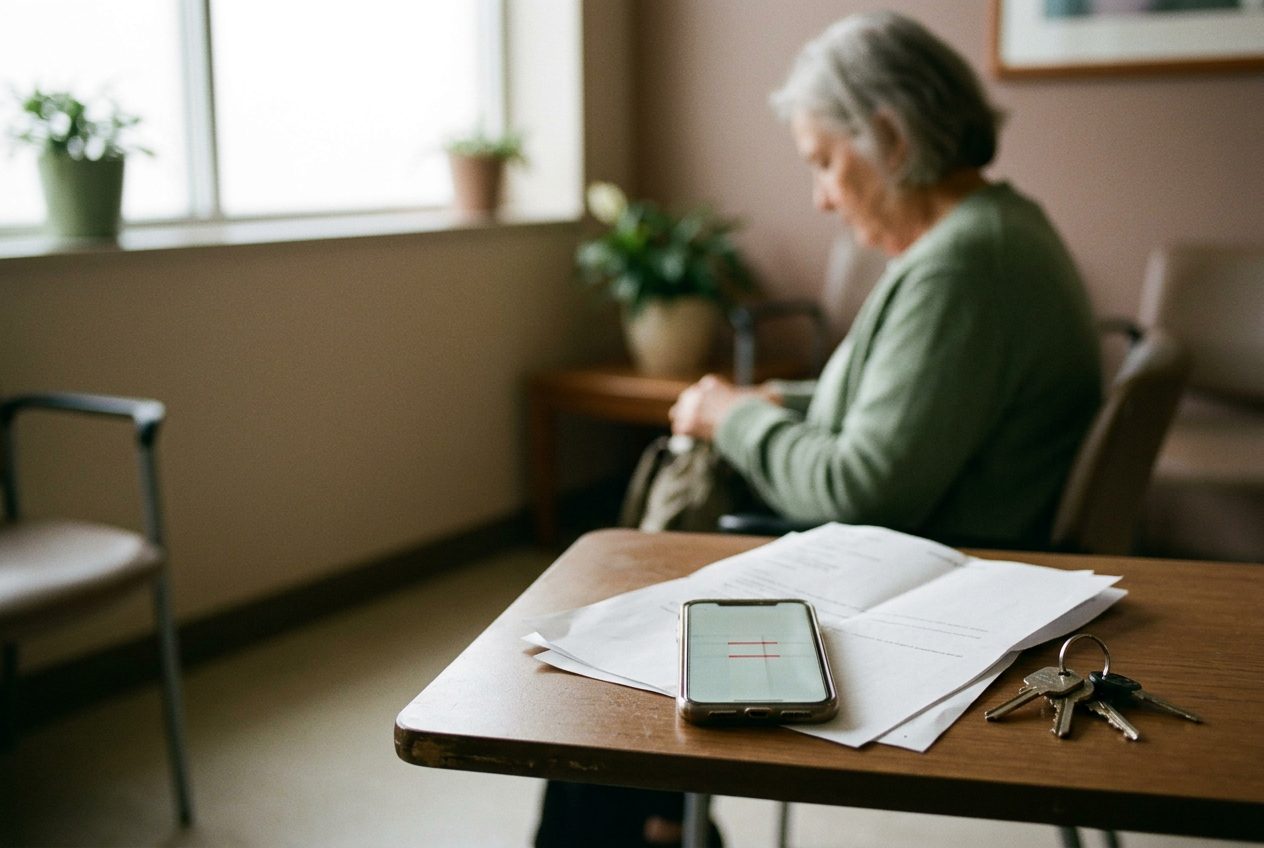
Maya woke after the examination with the printed portal page still folded in her bag. The line on it had read “EGD and possible biopsy.” Eight months of burning behind her breastbone and an unsettled stomach had led to that line, but when she first saw it, she thought she had been scheduled for surgery.
She had not.
EGD is short for esophagogastroduodenoscopy, usually called an upper endoscopy. A specialist passes a thin, flexible tube with a camera and light through the mouth to examine the esophagus, stomach, and duodenum, the first part of the small intestine. There is no skin incision. A biopsy means the specialist removes tiny tissue samples through the same tube for laboratory testing.
That is the short answer Maya needed before anything else: the camera and the samples were parts of one examination, not separate operations.
Two results, on different schedules
The specialist could tell Maya what the camera had shown soon after the EGD. The biopsy findings were not ready.
That split is easy to miss on a patient portal, which may show one procedure line without explaining that visual findings and microscopic findings arrive at different times. The specialist sees the digestive tract lining during the examination and can describe its appearance afterward, although sedation may make that conversation hard to remember. Maya also received written discharge information, and her companion listened.
A pathologist, a doctor who specializes in interpreting cells and tissue, must process and examine the biopsy samples under a microscope. That takes longer. Results often arrive within several days, though timing depends on the laboratory and whether the tissue needs more review.
Maya later received a portal notice that the pathology report was available. Her specialist explained how those findings fit with the camera examination and her earlier tests. The printed portal page stayed folded in her bag.
What an EGD can show
The camera gives the specialist a direct view of the upper digestive tract lining. It can reveal inflammation, ulcers, bleeding, narrowed areas, growths, and other visible changes. The specialist may take photographs or treat certain findings during the same procedure, depending on why the EGD was ordered and what appears during it.
Gastroesophageal reflux disease, or GERD, occurs when stomach contents repeatedly move into the esophagus and cause symptoms or complications. An upper endoscopy may show irritation or damage related to reflux. It may also reveal changes that need tissue testing.
A normal-looking esophagus does not rule out GERD. Many people with reflux symptoms have no visible injury during an EGD, which means the specialist has to consider the camera findings alongside the person’s symptoms and any other testing.
Celiac disease raises a different issue. It is an immune reaction to gluten, a protein in wheat and related grains, that can damage the small intestine. The duodenum may look normal through the camera even when injury is visible under a microscope.
This was the distinction Maya had been missing when she stared at the word biopsy. The camera showed the surface. The tissue samples could show changes too small for the camera to pick up.
The part that frightened her
A biopsy does not by itself mean the specialist suspects cancer.
During an upper endoscopy, specialists take tissue for many reasons. Esophagus samples can help identify inflammation or changes associated with long-standing reflux. Stomach tissue may be checked for infection or inflammation. Samples taken from different parts of the duodenum can show the pattern of intestinal injury associated with celiac disease.
The location matters even though the pieces are tiny. Each sample is placed in a container and sent to a laboratory. The pathologist then examines the tissue under a microscope and prepares a report for the specialist who ordered the examination.
For celiac disease, that report cannot be interpreted alone. The specialist also considers blood test results, symptoms, medical history, and what the person was eating before testing, because cutting back on gluten or removing it can change the findings. A general explainer cannot tell someone whether to alter their diet before an evaluation. That question belongs with the gastroenterology specialist managing the testing.
Maya had assumed that taking tissue required another operation. Once the office explained the endoscope’s instrument channel, the line on the printed portal page made more sense: small tools could pass through the tube while the camera remained in place.
Inside the procedure area
Before the EGD, a staff member reviewed Maya’s health history and placed an intravenous line, often called an IV, for medication. Equipment monitored her breathing, heart rate, and blood pressure. She lay on her side with a small guard between her teeth to protect her mouth and the endoscope.
Sedation means medication that reduces awareness and discomfort. Some people remain able to respond but remember little afterward. Others receive deeper sedation and are more fully asleep under the supervision of trained clinical staff. The plan depends on the setting, the person’s health, and the examination being performed.
After Maya was sedated, the specialist guided the endoscope through her mouth, behind her tongue, and into her esophagus. The tube did not enter her airway. Her breathing continued to be monitored while the specialist examined the esophagus, advanced into the stomach, and then viewed the duodenum.
Air or carbon dioxide may be introduced to open folds in the digestive tract lining so the specialist can see more clearly, while fluid may be rinsed or suctioned away if it blocks the view. The camera portion often takes about 10 to 20 minutes, though treatment or complicated findings can lengthen the examination.
The specialist passed small instruments through a channel inside the scope and collected several samples. No incision was made in Maya’s skin. Most people do not feel these samples being taken, and each piece was much smaller than Maya had pictured when she read the portal page.
Before an upper endoscopy
Preparation is not identical for everyone. Instructions depend on why the EGD was ordered, the person’s health, and the sedation plan.
Patients are generally told when to stop eating and drinking. The care team also reviews medications and explains whether another adult must drive the patient home. Maya’s specialist discussed her medicines, went over sedation, and confirmed that she had someone to take her home.
Those instructions can change for people who use blood thinners or diabetes medication, and they may also change when someone has allergies or heart and breathing conditions. The relevant directions are the ones provided by that person’s procedure team, not a general article.
By then, Maya understood more than she had when she printed the portal page. She still carried it with her. The words had not changed.
Recovery and risk
Recovery from sedation took Maya longer than the camera examination. She had temporary throat discomfort and bloating, both of which can occur after the scope passes through the mouth and gas is used to improve the view.
Her discharge paper explained which symptoms the team expected and which uncommon signs required prompt contact with the medical office or emergency care. Sedation can affect judgment and memory, which is one reason patients may need another adult to listen to the discharge discussion and drive them home.
Serious complications are uncommon. They can include a reaction to sedation, bleeding, a tear in the digestive tract, or material entering the airway. Personal risk depends on health history and what the specialist does during the procedure, so the specialist or anesthesia professional is the right person to explain that risk before consent.
This article cannot estimate one reader’s risk from an EGD or say which level of sedation is appropriate. It does not have access to that person’s medical conditions, medicines, allergies, or planned procedure.
What the examination cannot answer
An EGD does not examine most of the small intestine. It does not examine the colon.
It may not explain why someone has heartburn, nausea, pain, or trouble swallowing, and a normal examination does not make those symptoms unreal. Some conditions leave visible changes. Others do not. Biopsy results can add information, but they do not answer every question either.
This piece cannot tell a reader whether an EGD is needed or whether a biopsy confirms GERD or celiac disease. A gastroenterology specialist must connect the symptoms with the medication history, blood work, camera findings, and pathology report. The person who ordered the procedure can also explain why tissue was taken from a particular location.
Maya kept her printed portal page through the follow-up. Beside the line that had scared her, she wrote that the camera showed what the lining looked like and the samples showed what the tissue contained.
Questions people ask
Does a biopsy during an EGD mean the doctor suspects cancer?
No. Tissue may be taken during an upper endoscopy to look for inflammation, infection, reflux-related changes, or microscopic signs of celiac disease. The reason depends on where the samples came from and why the procedure was ordered. The specialist can explain that after reviewing both the procedure findings and the pathology report.
Will I feel the endoscope or the biopsy?
Sedation is commonly used to reduce awareness and discomfort. Most people do not feel the tiny tissue samples being removed from the digestive lining, though experiences vary with the sedation method and the person’s health.
The procedure team can explain the planned sedation and how breathing and circulation will be monitored. A general article cannot predict what one person will remember or feel.
Can a normal EGD rule out GERD or celiac disease?
No. An esophagus can look normal in someone who has GERD. The duodenum can also appear normal even when celiac-related injury is present under a microscope.
Biopsy findings, blood tests, symptoms, and treatment history may all affect the interpretation. The gastroenterology specialist has to place the EGD findings within the rest of the medical record.
When do results come back?
The specialist can usually discuss visible findings after the examination, although sedation may make the explanation difficult to remember. Biopsy samples require laboratory processing and often take several days, with longer waits possible if additional review is needed.
Maya’s final explanation came after the pathology report reached her portal. The original printed page was still folded in her bag.
One story a day
The story of the day, in your inbox
One health journey each morning — no advice, no alarm, just company for the road.